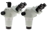
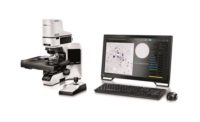
Stereomicroscopes have characteristics valuable in situations where three-dimensional observation, depth perception, and contrast are critical. These instruments are also essential when micromanipulation of the specimen is required in a large and comfortable working space. The wide field of view and variable magnification displayed by stereomicroscopes are also useful for construction of miniature industrial assemblies, or for biological research that requires careful manipulation of delicate and sensitive living organisms.
Considering the wide range of accessories currently available for stereomicroscope systems, this class of microscopes remains extremely useful in a multitude of applications. Stands and illuminating bases are available from all manufacturers, and can be adapted to virtually any working situation. There are a wide choice of objectives and eyepieces, enhanced with attachment lenses and coaxial illuminators fitted to the microscope as an intermediate tube. Working distances can range from three to five centimeters to as much as 20 centimeters in some models, allowing for a considerable amount of working room between the objective and specimen. For assembly, inspection, quality assurance, and scientific discovery, there is much to consider about stereomicroscopes.
Stereoscopic Vision
Our eyes and brain function together to produce what is referred to as stereoscopic vision, which provides spatial, three-dimensional images of the objects surrounding us. This is because the brain interprets two slightly different images received from each of our retinas. The average human eyes are separated by a distance of approximately 64-65 millimeters, and each eye perceives an object from a somewhat different viewpoint a few degrees apart from the other. When transmitted to the brain, images are fused together but still retain a high degree of depth perception, which is truly remarkable. The stereomicroscope takes advantage of this ability to perceive depth by transmitting twin images inclined by a small angle (usually between 10 and 12 degrees) to yield a true stereoscopic effect.
With historical roots in the 17th century, many new advances are continuing to take place in the world of stereomicroscopy. World firsts, such as a zoom ratio of 25:1, more advanced lighting methods, ever more sophisticated optics, and powerful data connections are capturing more and more applications for this inspection and analysis tool.
The first stereoscopic-style microscope having twin eyepieces and matching objectives was designed and built by Cherubin d’Orleans in 1671, but the instrument was actually a pseudo-stereoscopic system that achieved image erection only by the application of supplemental lenses. It wasn’t until more than 150 years later when Sir Charles Wheatstone wrote a treatise on binocular vision that enough interest was stimulated in stereomicroscopy to provide the impetus for further work.
In the early 1890s, Horatio S. Greenough, an American instrument designer, introduced a novel design that was to become the forerunner of modern stereomicroscopes. When he partnered with industry, engineers designed inverting prisms to produce an erect image. This design has withstood the test of time (and a large number of microscopists), and was a workhorse in medical and biological dissection throughout the 20th century. It is still a favorite for many specific applications.
Stereomicroscopes manufactured during the first half of the 20th century, or dissection microscopes as they were called, were much like traditional compound microscopes of the era. They were heavy, constructed mainly from brass, used prisms for image erection, and had simple lens systems consisting of one or two doublets. Working distance was inversely proportional to magnification, and was quite short at the highest available magnifications. These microscopes were employed primarily for dissection, because there were very few industrial applications involving small assemblies that required a microscope for examination. Even watchmakers used monocular loupes!
In 1959, a stereomicroscope was introduced with a cutting-edge advance: continuously variable, or zoom, magnification. This microscope was the first stereomicroscope without erecting prisms and was fashioned around the basic Greenough design. The microscope also featured: four first-surface mirrors with enhanced aluminum coatings, strategically positioned to perform the function of both inclination prisms and erecting prisms. In stereomicroscopy, erect images are useful because microscopists often must perform interactive manipulations on the specimen while under observation. Tasks such as dissection, micro-welding, industrial assembly, or microinjection of oocytes are more conveniently conducted when the specimen has the same physical orientation on the microscope stage as it does when viewed through the eyepieces. Also, the study of true spatial relationships between specimen features is aided by a natural, erect image.
Stereomicroscopes of the Greenough design have been very popular in the assembly and inspection of small components, particularly in electronics. The working distance affords the opportunity to place tools under the microscope, for tasks such as soldering of small electronic components on printed circuit boards (PCBs).
For more complex configurations, a modular design is required. Higher end stereomicroscopes use another design known as the CMO (common main objective) stereomicroscope because a single large objective lens at the bottom of the body accumulated light from the specimen for the left and right channels. The greatest design feature and practical advantage of a common main objective stereomicroscope, as with most modern microscopes, is the infinity optical system. A collimated light pathway, with two parallel axes for the channels, exists between the objective and removable head/observation tube assembly. This allows the effortless introduction of accessories, such as beamsplitters, coaxial episcopic illuminators, photo or digital video intermediate tubes, drawing tubes, eyelevel risers, and image transfer tubes into the space between the microscope body and head. It is also possible to place these accessories in the space between the objective and zoom body, although this is rarely done in practice. Because the optical system produces a parallel bundle of light rays between the body and microscope head, the added accessories do not introduce significant aberrations or shift the position of images observed in the microscope.
Common main objective microscopes are generally used for more complex applications requiring high resolution with advanced optical and illumination accessories. The wide spectrum of accessories available for these microscopes lends to their strength in the research arena. In many industrial situations, Greenough microscopes are likely to be found in production lines, while common main objective microscopes are limited to the research and development laboratories. Another consideration is the economics of microscope purchase, especially on a large scale. Common main objective stereomicroscopes can cost several times more than a Greenough microscope, which is a chief consideration for manufacturers who may require tens to hundreds of microscopes. However, there are exceptions. If a common main objective microscope is the better tool for a job, the true cost of ownership may be lower in the end.
21st Century Advances
Many new features are shifting stereomicroscope usage and value. A new stereoscope design dynamically changes the distance between the two optical axes as the zoom factor is changed. Change in optical axis distance enables maximization of light entry into the optical system at every magnification. The result is an uncompromised, large zoom range, up to 25:1, with high resolution in both eye paths, and minimal aberrations over the entire zoom-range. Furthermore, this breakthrough in optical design enables all these desirable features to be housed in a compact zoom body.
Auto link zoom (ALZ) automatically adjusts the zoom factor to maintain the same field of view when switching objective lenses. This function enables seamless switching between whole organism imaging at low magnifications and detailed imaging at high magnifications.
Oblique coherent contrast (OCC) is a form of oblique lighting method. Compared to conventional diascopic illumination that illuminates directly from below, OCC illumination applies coherent light to samples in a diagonal direction, giving contrast to colorless and transparent sample structures, such as plastics and glass materials.


