10 Common Microscope Challenges
Today’s most common microscope challenges are solved with advanced digital microscopy.


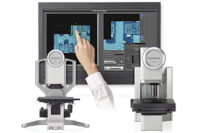
Users of conventional optical and digital microscopes face a long list of challenges when trying to perform advanced imaging and measuring operations. With today’s newest digital microscopy technology, these challenges can be met. Made possible by the combination of time-tested optics and advanced digital imaging technology, today’s latest digital microscopes allow even first-time users to immediately produce superior images and highly reliable results.
Advanced digital microscopes allow operators of conventional optical and digital microscopes to overcome a variety of challenges related to observation, image capturing, and measurement. Featured here are 10 common conventional microscope challenges and 10 solutions made possible by current advances in digital microscopy.
CHALLENGE 1: Low contrast and glare make it hard to see specific sample details.
In many imaging cases, a sample with low contrast cannot be clearly observed. In addition, some areas on a sample image can be too bright or too dark due to the sample material, texture or color, which can make it difficult to clearly observe surface conditions. To eliminate those inconveniences, illumination needs to be carefully adjusted, which can require considerable time.
SOLUTION: The combination of high-quality optics and advanced digital image processing allows optimized imaging without complex adjustment.
Today’s newest digital microscopes combine high-quality optics with advanced digital image processing features to allow clear observation of previously difficult surface conditions. For example, digital microscopes can offer an HDR (High Dynamic Range) function that combines several images taken at different exposures, enabling clear observation even with low-contrast images. With HDR, samples that previously may have required multiple pieces of equipment for precise observation can be clearly observed with a single system.
CHALLENGE 2: It is difficult to view an entire large sample at high resolution.
With conventional microscopes, it is often difficult to view an entire sample at high resolution. As magnification is increased, the observation field becomes narrower, and it becomes very hard to obtain an overall image of the sample.
SOLUTION: With new digital microscope technology, there is no longer such a thing as “outside the field of view.”
With high-quality panoramic imaging, today’s latest digital microscopes can automatically stitch images into one seamless view (Fig. 1) by simply moving the stage. As the stage moves, the panoramic imaging system automatically stitches images into a large single field of view, in real time. Where conventional microscopes reduce field area with increases in magnification, digital panoramic imaging maintains the original field while delivering close-up clarity.
CHALLENGE 3: All areas of an uneven sample surface are hard to view in clear focus at the same time.
With conventional microscopes, the more the operator increases the magnification, the shallower the depth of focus can become, making it more difficult to achieve clear focus on the entire sample.
SOLUTION: Advanced digital microscopes make it possible to obtain a clear, in-focus image of an entire sample with one simple click—no matter how uneven the surface.
With the EFI (Extended Focal Image) function, several images are taken while the point of focus is moved up and down. From these images, the areas where the sample was in focus are combined into one image where the whole sample is in focus, allowing the precise inspection of uneven surfaces.
CHALLENGE 4: Sample features need to be determined, characterized and measured in 3-D.
Using an optical microscope, it is difficult to determine the exact features of a dimensional sample based on a 2-D image. In many cases, the operator has no alternative but to make their best estimate as to height differences in a sample or whether sample unevenness is concave or convex (Fig. 2).
SOLUTION: With one click, today’s digital microscopes can image samples in three dimensions, allowing examination from any angle and a view of the sample as it actually is.
With detailed 3-D images, sample features or unevenness can be easily viewed and measured (Fig. 3). Height differences and volume can also be measured, making it easier to accurately analyze the sample. Three-dimensional imaging is simple and fast.
CHALLENGE 5: Microscope operators with minimal experience need to perform advanced tasks.
With traditional optical microscopes, switching between observation modes can require a variety of complicated operations and adjustments. Without considerable knowledge and experience, it can be difficult to ascertain the specific image characteristics one needs for the task at hand. Changing the observation mode can often require adjustment of aperture stop and illumination, the insertion of special filters, etc.
SOLUTION: Simply select your optimal image, and your observation mode will match your selection.
Advanced digital microscopy has changed microscope operation flow dramatically. Simply select your optimal image from a list of captured image choices, and your observation mode will be adjusted based on your selection.
CHALLENGE 6: Reproducible measurement results are needed from multiple operators.
With conventional microscopes that require many fine adjustments and settings, it can be difficult for multiple users to conduct observations under the same conditions. Observation conditions may vary depending on the operator and their particular method, which can cause major differences in the images observed. This can cause problems when conducting R&D, quality assurance, or material testing. In addition, if a digital camera is mounted to a microscope but the microscope is not linked to image processing software, image processing can take multiple time-consuming steps (the microscope and software are operated separately). Moreover, if the magnification settings of the software are not correct, measurements may be conducted with incorrect calibration values from user to user.
SOLUTION: Hardware and software designed to work as one.
With a fully digital microscope, all image acquisition and observation conditions, including stage coordinates, observation method, calibration data, etc., can be saved and recalled at any time. Any operator can easily repeat any inspection and measurement method, ensuring observation under the exact same conditions and settings. All image capture conditions can be recalled with one simple click, and all operations are simply and speedily controlled by software that operates seamlessly with the system’s mainframe.
CHALLENGE 7: High-resolution imaging is needed from a digital microscope.
Many previous digital microscopes provide ease of use but do not deliver the same high resolution as an optical microscope. In fact, many users divide their microscope usage between the two depending on their application. This wastes large amounts of time and does not allow continuity of observation between digital and optical images. In addition, when conducting observations at a high magnification using some digital microscopes, the impact of vibration can cause blurring. In many cases, this can make it impossible to observe fine sample details.
SOLUTION: Today’s newest digital microscopes provide both high-quality imaging and easy operation, thanks to advanced optics and a stable frame.
Advanced digital microscopes feature objective lenses that combine high NA, long working distances, and evenness of light intensity to minimize glare and allow true color reproduction. Flare and distortion are gone, unheard of with traditional digital microscopes. These new digital microscopes also feature sturdy, high-rigidity frames with a low center of gravity, specifically designed to absorb the impact of vibrations. Stable observations can be conducted at magnifications as high as 9,000x.
CHALLENGE 8: Guaranteed measurement accuracy is needed from a digital microscope.
Traditional digital microscopes offer significant depth of focus. Depending on the point of focus, however, the size of the captured image may vary. If the point of focus varies even slightly under the same magnification, then variation in measurement results can occur—there are no clear guarantees regarding the reliability of traditional digital microscopes. Operators therefore have no way of being assured of the scope of error of the equipment they are using.
SOLUTION: With new digital microscope technology, many factors of uncertainty are removed, and measurement results can be guaranteed.
Today’s newest digital microscopes utilize the same telecentric optics found in measuring instruments, eliminating variation in measurement results. Even if the point of focus is changed, there is no change in the size of the target of observation.
CHALLENGE 9: Different
observation modes require different lens setups.
Traditional digital microscopes require time-consuming lens replacement in accordance with the illumination method being used. Every time the lens is changed, it is also necessary to remove and reattach the camera and cables, which requires even more time and effort.
SOLUTION: With an advanced digital microscope, just one click can change your observation method. Any operator, beginner to expert, can conduct the same level of observation.
Advanced digital microscopes can switch between observation methods with one simple click, efficiently finding the appropriate observation method for the sample in question. Aperture stop adjustment, illumination, and special filters are automatically optimized so that any operator, regardless of skill level, can conduct appropriate observations.
CHALLENGE 10: Magnification changes require manual
calibration.
Many traditional digital microscopes require troublesome calibration settings to be performed every time the magnification is changed. If images or measurements are taken using incorrect calibration settings, the magnification indication and measurement values will also be incorrect, requiring the whole process to be performed again.
SOLUTION: Automatic magnification recognition eliminates calibration errors.
Digital microscopes (Fig. 4) offer automatic magnification recognition, with a coded zoom system and a coded nosepiece that automatically tell the system what objective is in use. When you measure something on the image, your calibration is always correct.
Thanks to high-quality optics and advanced digital technology, today’s latest digital microscopes deliver efficient observation, intuitive magnifying operation, a variety of observation methods, and reproducibility. Various image capturing methods provide easy, intuitive operation—as simple as using a smartphone or tablet. Options include EFI and 3-D imaging, wide area image capturing, movie capturing, and programmed image capturing. Measurement is backed by guaranteed accuracy and repeatability, automatic calibration, and reproducibility self-check. Advanced reporting systems allow easy sharing of measurement and analysis results.
Looking for a reprint of this article?
From high-res PDFs to custom plaques, order your copy today!


